Tripier Flap with Lateral Periosteal Strip for Lower Eyelid Ectropion/Retraction
Transcript
I prefer Tripier flaps when I am trying to address inferior displacement of the lateral canthus and lateral lower eyelid retraction/ectropion. The flap is long, and it is likely that much of it is not perfused and is acting more like a skin graft. However, the area by the base of the flap is the most important area that is addressed, and this area is likely perfused. Transposition of the flap results in lengthening of the lower lid but also raises the lateral canthus.
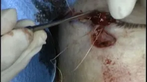
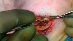
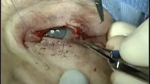
This is Richard Allen at oculosurg.com. This video demonstrates the use of a Tripier flap with a lateral periosteal strip to repair lower eyelid retraction/ectropion. The patient has had previous attempts to repair a predominantly lateral lower lid retraction/ectropion. A blepharoplasty has been marked which retains a lateral pedicle. The 15 blade is used to make an incision along the marking extending to the lateral canthus, retaining a lateral pedicle. The myocutaneous flap is then raised by dissecting between the orbicularis muscle and the orbital septum. I try to use only scissors without cautery. A Frost suture is placed through the lower eyelid. A subciliary incision is then made with the 15 blade and dissection is carried out inferiorly between the orbicularis muscle and the orbital septum. Dissection planes are not as obvious due to scar from the previous operations. The goal is to make the lower eyelid freely mobile by releasing all scar. The transposition of the flap is then demonstrated. A lateral inferior cantholysis is then performed. The freer periosteal elevator is then used to expose the periosteum of the lateral orbital rim. A 15 blade is then used to make an incision through the periosteum of the lateral orbital rim so that a periosteal flap can be raised. This is performed to give extra strength to the lateral canthoplasty. The periosteal flap is raised with the freer periosteal elevator. Placement of the periosteal flap to overlap the lateral tarsus is demonstrated. The mucocutaneous junction of the lateral tarsus is excised. A 5-0 Vicryl suture is then placed in a mattress fashion so that the periosteal strip will be sutured to the anterior surface of the lateral tarsus. Tying the suture places the canthus in good position and stabilizes the lower eyelid. The Tripier flap is then transposed. There appears to be tension laterally, therefore, the lateral cheek is engaged with a 4-0 Vicryl suture which then engages the periosteum of the lateral orbital rim. Tying the suture elevates the cheek, relieving some of the tension. The Tripier flap is then suture into position with a combination of interrupted and running 5-0 fast absorbing sutures. Although I rarely trim flaps, the end of the pedicle will be trimmed in this case as it appears redundant. Additional sutures are placed. The lateral canthus is then reformed. The upper eyelid incision is then closed. The lower eyelid appears to be in good position. The Frost suture will be taped to the patient’s forehead, and the patient will return in one week for patch and Frost suture removal.