Medial Pillar Tarsorrhaphy as a Medial Canthoplasty
Transcript
There are many techniques for medial canthoplasty, all with their advantages and disadvantages. In this video, a pillar tarsorrhaphy is used to help support the medial lower eyelid. This is a straight-forward procedure and the traction on the eyelid is medial, posterior, and superior. The pillar is also barely noticeable when it is placed medially.
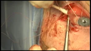
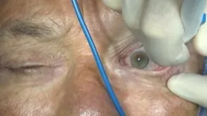
This is Richard Allen at oculosurg.com. This video demonstrates medial lower eyelid elevation with a pillar tarsorrhaphy. This patient has a history of a facial nerve palsy and is status post multiple procedures including retractor disinsertion with placement of a spacer to help elevate the lower lid. Unfortunately, the medial lower eyelid is still inferiorly displaced with exposure keratopathy. A 4-0 Silk suture is placed through the upper and lower eyelid margin to help provide traction during the case. Attention is directed to the lower eyelid where the needle tip cautery is used to make a transconjunctival incision inferior to the inferior boarder of the tarsus. Steven’s scissors are then used to release any scar along the medial lower eyelid. Traction on the silk suture demonstrates that the lower eyelid is not completely released. Additional blunt dissection is performed and the eyelid appears to be able to be mobilized. The pillar tarsorrhaphy is then planned and a shallow portion of the lower eyelid margin is deepithelialized with a 15 blade and Westcott scissors. This needs to be relatively shallow to avoid disruption of the canaliculus, but realistically in this case the lower canaliculus is likely not functioning. The upper eyelid is then everted and the patient’s previously placed weight is visible. The tongue of tarsus and conjunctiva will need to be medial to the weight and the weight should not be disturbed. The 15 blade is then used to make an incision inferior to the superior border of the tarsus. Westcott scissors are then used to develop the pillar. This includes tarsus, conjunctiva, and likely some Muller muscle. I would tend toward making the pillar too short, as this could be lengthened postoperatively if needed. The pillar is then engaged with a double-armed 5 or 6-0 Vicryl suture. Each arm of the suture is then placed through the area of deepithelialization on the lower eyelid, again trying to avoid the canaliculus. The sutures are then tied. Elevation of the medial portion of the lower eyelid is demonstrated. Inspecting pre- and post-operative photos demonstrates the effectiveness of the procedure and also that the pillar itself is relatively inconspicuous.