Placement of Bioengineered Spacer Graft for Lower Eyelid Retraction
Transcript
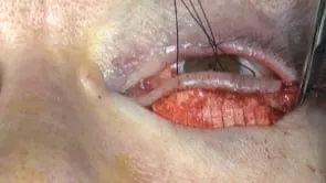
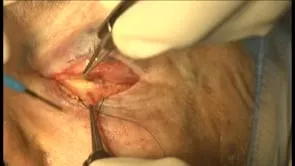
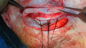
This video describes the placement of a bio-engineered spacer graft for lower eyelid retraction repair. The authors are doctors, Craig Sis, Kenneth Cahill, Jill Foster and David Hulk. Traction sutures are helpful, especially if the surgeries performed without a atomy for concomitant lateral lid tightening. Additional local anesthetic can be injected to help identify and separate the tissue layers. The conjunctival incision is marked one to two millimeters below the inferior tarsal margin. It extends from the edge of the care uncle to the lateral canthal area. Conjunctival traction sutures are helpful. The incision passes through the lower lid retractors so that resistance to upward distension of the lid margin is eliminated. The lower lid fat pads should be made visible. Processed porcine collagen is hydrated for at least 20 minutes. It is trimmed to the desired size. It should be approximately three millimeters in vertical height for every millimeter of desired lid margin elevation inferiorly, it is sutured to the recessed lower lid retractors with five or six oh. Vicryl Care should be taken to bury the knots. Sutures can be interrupted or run. The edges of the conjunctiva should remain over the edge of the spacer graft. It is best to minimize or avoid leaving suture loops exposed. The superior edge of the spacer is sutured to the inferior tarsal margin. Sutures can be interrupted or run. The vertical position of the lid margin should be slightly overcorrected. At the conclusion it is best to minimize or avoid leaving suture loops exposed. Frost sutures or temporary suture tar sophis can be left in place postoperatively if desired.