Placement of Upper Eyelid Weight with Existing Pillar Tarsorrhaphy
Transcript
I use pillar tarsorrhaphies liberally. I believe that there are numerous advantages: the eye can be examined easily, the lid margin is not disrupted, and it can be easily opened in stages with just topical anesthetic. The only disadvantage is that it does not provide a water tight closure, which is sometimes needed in cases of significant keratopathy. I will often place the pillars, then do other surgeries that will be needed especially in cases of facial nerve palsy. In this case, the pillars have been placed previously, and an upper lid weight will be placed while the pillars are in. The pillars can then be opened slowly at a later date.
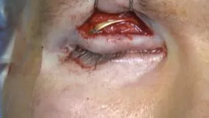
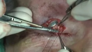
This is Richard Allen at oculosurg.com. The video demonstrates placement of an upper eyelid weight in a patient who already had a previously placed pillar tarsorrhaphy. The pillar tarsorrhaphy is demonstrated. This patient has a facial nerve palsy and a neurotrophic cornea. A lid crease has been marked. The 15 blade is used to make an incision along the lid crease marking. Dissection is carried out to the underlying orbital septum, the orbital septum is opened, and the preaponeurotic fat is dissected from the underlying levator aponeurosis. A thermal cautery is used to disinsert the levator aponeurosis from the anterior surface of the tarsus, and dissection is carried out between the levator aponeurosis and the underlying Muller muscle. A 1.2 gram platinum weight is placed between the levator aponeurosis and Muller muscle. The inferior holes on the weight are sutured to the superior boarder of the tarsus with 6-0 Prolene sutures. The cut end of the levator aponeurosis is sutured to the edge of the pretarsal orbicularis with interrupted 7-0 Vicryl suture. This helps in formation of the lid crease and recesses the levator just a little. The lid crease incision is then closed with interrupted 5-0 fast absorbing sutures.