Combined Frontalis Flap and Levator Resection
Transcript
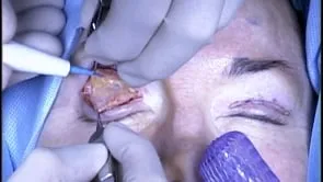
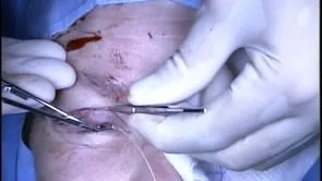
This is Richard Allen at oculosurg.com. The video demonstrates a combined frontalis flap and levator resection for poor levator function, severe congenital ptosis. The proposed lid crease has been marked, and a 15 blade is used to make an incision along the marking. Dissection is then carried out through the orbicularis muscle to the underlying orbital septum. The orbital septum is exposed and dissection is performed through the orbital septum to identify the underlying preaponeurotic fat. The septum is thick in children. The preaponeurotic fat is then dissected from the underlying levator aponeurosis and muscle. Examination of the muscle shows that it is infiltrated with fat, consistent with poor levator function, severe congenital ptosis. Dissection is then carried out superiorly along the surface of the preaponeurotic fat with Westcott scissors to the arcus marginalis and periosteum of the superior orbital rim. Dissection is then carried out along the anterior surface of the brow fat, posterior to the frontalis muscle. This is performed along the horizontal extent of the superior orbital rim, being aware of the supraorbital neurovascular bundle medially. This dissection is carried out about 2 cm superior to the superior orbital rim. There can be some bleeding that can be controlled with cautery. Dissection is then redirected along the anterior surface of the frontalis muscle, between the subcutaneous fat and the frontalis muscle. As can be seen, the muscle fibers are identified, and I am initially beneath the muscle fibers, and then I need to redirect my dissection anteriorly. This dissection is carried out approximated 2 cm superiorly. It is not uncommon to see brow hair follicles and it is important to not button hole through the skin. The mobilized flap is demonstrated. The preaponeurotic fat is repositioned into the orbit. The positioning of the flap is again demonstrated. The levator aponeurosis is then disinserted from the anterior surface of the tarsus. A combined levator resection will be performed to help create a lid crease and prevent “lid-popping”. Dissection is carried out between the levator aponeurosis and the Muller muscle. A double-armed 5-0 nylon suture on a spatula needle is placed partial thickness through the anterior surface of the tarsus at the level of the medial limbus. Each arm is then placed through the levator aponeurosis followed by the frontalis flap. Tightening the suture should place the eyelid margin at the superior limbus. An additional nylon suture is placed temporally, engaging the levator aponeurosis followed by the frontalis flap. Again, tightening the sutures checks the height and contour of the eyelid. The sutures are then tied. The end of the levator aponeurosis is then identified and the redundant portion is excised. The edge of the levator aponeurosis will then be utilized to aid in lid crease formation. The incision is closed with 5-0 fast absorbing sutures which engage the edge of the levator aponeurosis. Antibiotic ointment is placed over the incision and into the eye. A double eye pad will be placed for 1-2 days and the patient will follow up in one week.