MMCR with Tarsectomy for Congenital Ptosis
Transcript
Treatment of congenital ptosis can be performed with multiple different procedures. For minor amounts of ptosis (2 mm or less) that respond to phenylephrine, an MMCR is an effective procedure to use. However, if there is an incomplete response to the phenylephrine, I would consider adding a tarsectomy to the 9 mm MMCR. The amount of the tarsectomy is twice the amount of the incomplete response, up to 2 mm of tarsectomy. I would not do more than 2 mm of tarsectomy. For example, if a patient has 2 mm of ptosis and phenylephrine lifts the eyelid 1.5 mm, a 1 mm tarsectomy would be added. If the patient has 2 mm of ptosis and phenylephrine lifts the eyelid 1 mm, a 2 mm tarsectomy would be added.
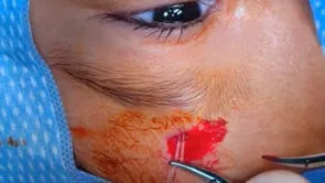
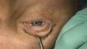
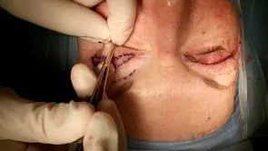
This is Richard Allen at oculosurg.com. This video demonstrates repair of congenital ptosis using a Muller muscle-conjunctival resection (MMCR) and 1 mm tarsectomy. I will use this procedure for mild amounts of ptosis that do not respond completely to phenylephrine. A 4-0 silk suture is placed through the eyelid for traction during the case. The eyelid is everted over a Desmarres retractor. The superior border of the tarsus is marked with the needle tip cautery or a marking pen denoting the middle one-third of the tarsus. In this case, the patient has 1.5 mm of ptosis and phenylephrine raised the eyelid 1 mm. Therefore, using the 2 to 1 ratio, a 1 mm tarsectomy will be added to the MMCR. Markings are then made 4 mm superior to the markings on the superior border of the tarsus. Markings are then made 1 mm inferior to the superior border of the tarsus for the tarsectomy. Toothed forceps are used to grasp the markings on the conjunctiva and the Putterman clamp is placed over the tissue to the tarsal markings. A 6-0 Plain gut suture is then placed in a running mattress fashion under the clamp. I start laterally and move medially, then return to the lateral area. This will place the knot lateral, which is less irritating for the patient. You could also bury then knot under the conjunctiva or place the suture transcutaneously. A 15 blade is then used to excise the tissue in a metal on metal fashion. I like to pull on the sutures to ensure that the 15 blade did not accidentally cut the suture. The suture is then tied. Inspecting the resected tissue, one can see the excised conjunctiva, Muller muscle, and small amount of tarsus. Antibiotic ointment is placed in the eye and used three times a day until the patient follows up in approximately one week.