Utilization of Fibrin Sealant for Wound Closure in Müller's Muscle Conjunctival Resection Ptosis Repair
Transcript
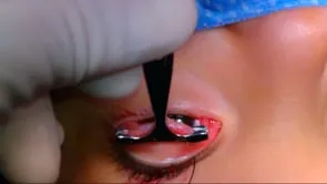
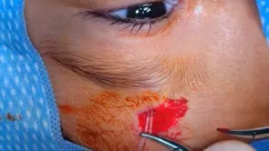
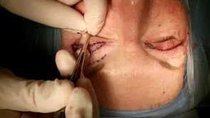
Utilization of fibrin sealant for wound closure in Mueller's muscle conjunctival resection, ptosis repair, authored by doctors Marsha Kavanaugh, Craig Siz, Jill Foster, Julian Perry, and David Hulk. A four oh silk traction suture is placed partial thickness through the eyelid. The eyelid is averted over a das more retractor, wrapping the traction suture around the handle and stabilizing it with a hemostat. Additional local anesthetic is injected into the conjunctiva. The amount of desired resection is measured using calipers and marked on the conjunctival surface with monopolar cautery. A putterman clamp is used to ate the conjunctiva and Mueller's muscle at the marked location. Two locking 0.5 forceps are placed medially and laterally to stabilize the tissues. A 15 barred Parker blade is used to inside the conjunctiva and Mueller's muscle just inferior to the clamp Monopolar. Cautery is used to achieve hemostasis. It is essential that complete hemostasis is achieved because excess blood products in the wound may interact with the fibrin sealant, thereby reducing its efficacy. The wound is re approximated using 0.5 or 0.3 forceps and Hartman hemostat clamps. The fibrin sealant is prepared according to the manufacturer's directions and applied to the wound here is taken to ensure all of the wound is adequately covered. The sealant is allowed to dry for approximately two minutes. The DeMar retractor is removed from the upper lid. Any excess sealant should be excised with wescott scissors. The clamps are then removed without disrupting the reapproximate tissues. Note how the wound is well approximated via the tensile strength of the fibrin sealant. The wound should be carefully inspected for any areas of micro dehisces and if necessary, re glue. The eyelid is placed into his normal position and the traction suture is removed.