Evisceration of an Eye Containing Silicone Oil
Transcript
My preference for performing eviscerations over enucleations has increased significantly over the years. Of course, it is mandatory to ensure that the eye does not have evidence of an intraocular malignancy. This can be done with direct inspection if there is a view to the retina, or an ultrasound or other imaging can be used. I used to think that eyes with silicone oil should only have an enucleation. However, I now routinely perform eviscerations on blind painful eyes with silicone oil. I believe that the advantage over an enucleation is that these eyes have often undergone multiple surgeries and the conjunctiva has been compromised and is thin, which increased your risk of implant exposure after enucleation.
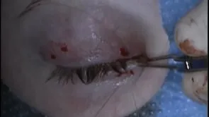
This is Richard Allen at oculosurg.com. This video demonstrates an evisceration in a patient who has silicone oil in the eye. I used to think this was a contraindication to an evisceration, but I no longer believe this. A 360 degree peritomy is performed with Westcott scissors. You can see that the conjunctiva is adherent to the globe from previous surgeries. A 15 blade is used to make a stab incision just posterior to the limbus. Westcott scissors are then used to perform a 360 degree incision just posterior to the limbus. Minimal pressure should be placed on the eye as you would like to remove the oil without it contacting the ocular surface. The suction follows the dissection to collect any oil. The suction is then placed directly in the eye to remove the oil.
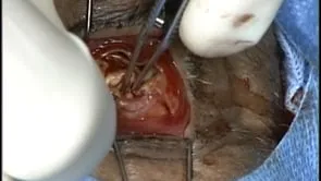
This can require the suction to be turned up as the oil is somewhat viscous. I like to use a disposable suction if possible as the oil can sometimes be difficult to clean from the instruments. Minimal irrigation is used to aid in floating the oil anteriorly. An evisceration spoon is then placed in the suprachoroidal space and the retina and choroid are removed and sent to the pathologist. Additional irrigation is performed with saline. The inside of the sclera is then scrubbed with dehydrated alcohol. The purpose of this is to denature any residual retinal or choroidal tissue. I will soak the sclera with the dehydrated alcohol. An incision is made in the posterior sclera. The optic nerve is transected. The intraconal space is demonstrated. Relaxing incisions are made 180 degrees away from each other in the anterior sclera. These are placed perpendicular to the posterior sclera incision. In this case, a porous implant will be placed. This is a relatively young patient and I was worried about potential migration of a non-porous implant. The implant inserter is used to place the implant in the intraconal space, posterior to posterior sclera. The residual optic nerve attached to the posterior sclera is removed. The posterior sclera is then closed over the implant with interrupted 4-0 Vicryl sutures. The anterior sclera is then closed with a running 4-0 Vicryl suture to imbricate one edge of the anterior sclera over the other. The conjunctiva is then closed with a running 7-0 Vicryl suture. A conformer will be placed and I will sometimes place a temporary suture tarsorrhaphy. Antibiotic will be placed with an eye patch which will be removed when the patient follows up in one week.