Free Composite Graft in Lower Eyelid Reconstruction
Transcript
The use of a composite graft for repair of full-thickness defects has gained some attention lately. I was always taught that since the graft had no blood supply, it would die. I have used this procedure sparingly in special situations, and have been pleasantly surprised with the results. My feeling is that the graft likely provide a template for granulation, more than anything else.
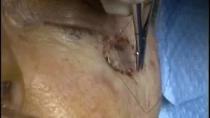
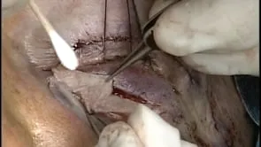
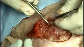
This is Richard Allen at oculosurg.com. This video demonstrates the use of a composite graft to repair a 60% full-thickness defect of the lower eyelid. The patient has poor vision on the contralateral side and relies upon the left eye for vision. The defect is measured on tension and the contralateral lower eyelid is marked. By transferring 30% of the contralateral lower lid to the left lower lid, the 60% defect is converted into a 30% defect, which can then be closed primarily. Both the height and width of the defect is measured. The composite graft is then harvested with a 15 blade and Westcott scissors. The donor site is then closed in the standard fashion for a wedge excision with 5-0 Vicryl suture through the anterior surface of the tarsus followed by 7-0 Vicryl suture placed in a vertical mattress fashion at the eyelid margin. These are placed at the level of the Meibomian gland orifices and lash follicles. The skin is the closed with interrupted 5-0 fast absorbing sutures. Attention is then directed to the left side where the composite graft is sutured to the adjacent eyelid. This is performed similarly to the wedge closure, with 5-0 Vicryl suture through the anterior surface of the tarsus. The eyelid margin is then repaired on each side of the composite graft with 7-0 Vicryl suture placed in a vertical mattress fashion at the level of the Meibomian gland orifices and lash follicles. The skin is then sutured with interrupted 5-0 fast absorbing sutures. I was always taught that this procedure should not work and the graft would die. Realistically, I believe the composite graft provides a template for granulation. Antibiotic ointment is placed over the repair three times per day for one week and the patient will follow up for reevaluation in one week. Inspecting the repair 2 months postoperatively shows the results on each side. Donor site is healed well, while the composite graft area shows some alteration of the eyelid architecture.