Anterior Lamellar Recession Lower Lid
Transcript
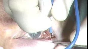
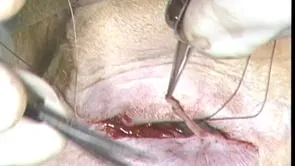
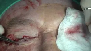
This is Richard Allen at the University of Iowa. This video demonstrates repair of segmental entropion of the right lower lid using an anterior lamellar recession. A 15 blade is used to make an incision along the eyelid margin at the grey line to separate the anterior and posterior lamella along the area of the entropion. Westcott scissors are used to further separate the pretarsal orbicularis muscle from the entire height of the anterior surface of the tarsus. The patient has some residual lashes along the anterior lamella that are excised with the Westcott scissors. These lashes could be retained if desired, but my preference is to usually excise them, depending on the patient. The anterior lamella is then recessed with a 6-0 vicryl mattress suture. The suture is placed approximately 1 mm inferior to the superior border of the anterior lamella, followed by a partial thickness pass through the anterior surface of the tarsus 2-3 mm inferior to the superior border of the posterior lamella, followed by passage through the anterior lamella. Tying the suture results in recession of the anterior lamella 1-2 mm. Three to four mattress sutures are placed. At the conclusion of the case, the anterior lamella is recessed. The superior defect along the posterior lamella is allowed to granulate. Antibiotic ointment is placed over the repair three times a day and the patient returns in one week for suture removal.