Skin Redrape with Canthal Plication for BPES
Transcript
Addressing the medial canthus in blepharophimosis, ptosis, epicanthus inversus syndrome (BPES) can be performed with a number of techniques. I had favored the four-flap (Y to V, double Z-plasty; “jumping man”) technique for many years. I was introduced to the medial canthal tendon plication/lower lid skin redraping technique, and now I prefer this technique. I think it makes sense in that the canthal abnormalities (telecanthus and epicanthus inversus) are addressed.
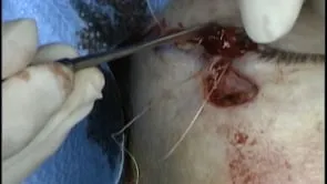
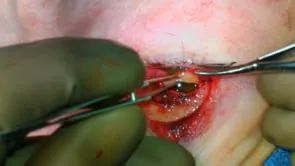
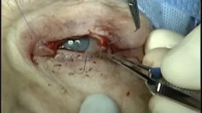
This is Richard Allen at oculosurg.com. This video demonstrates lower eyelid skin redraping with medial canthal tendon plication for blepharophimosis, ptosis, epicanthus inversus syndrome. I now do this procedure rather than the four-flap technique of a Y to V with double Z-plasty for this problem. I think it makes sense since the canthal issue is an epicanthus inversus with telecanthus. A marking has been made extending from the proposed medial location of the canthus to the medial canthal angle, and then extends an equal distance along the lower eyelid. A 15 blade is used to make an incision along the marking. This is a bilateral procedure and each side will be performed at the same time. Blunt dissection is then performed medially to expose the anterior limb of the medial canthal tendon. Wide undermining is performed posterior to the orbicularis muscle so that the tissue can be redraped. The medial canthal tendon is then exposed. A 4-0 Vicryl suture on a P-2 needle is used to engage the area of the insertion of the medial canthal tendon at the periosteum medially. The suture then engages the insertion of the medial canthal tendon at the medial canthal angle. Tying the sutures results in plication of the medial canthal tendon and transposition of the medial canthal angle medially. The same procedure is then performed on the opposite side. Stevens scissors are used to bluntly dissect to expose the medial canthal tendon. The insertion of the medial canthal tendon medially at the periosteum is engaged with the 4-0 Vicryl suture. The suture then engages the insertion of the medial canthal tendon at the medial canthal angle. Tying the sutures results in plication of the medial canthal tendon and transposition of the medial canthus medially. The lower eyelid skin is then redistributed along the newly mobilized superior edge of the incision. This is sutured into position with deep interrupted 6-0 Vicryl sutures. The skin is then closed with interrupted superficial 7-0 Vicryl sutures. Erythromycin ophthalmic ointment will be applied over the repair three time per day for one week, and the patient will follow up for reevaluation in approximately one week.