Harvesting a Sural Nerve Graft
Transcript
As corneal neurotization is becoming more common, harvesting of the sural nerve for the nerve graft is a useful procedure to learn for the oculoplastic surgeon. The anatomy is relatively straight-forward. Although the patients may have numbness in the area of the lower lateral leg, lateral heel, ankle, and dorsal foot, this often improves with time.
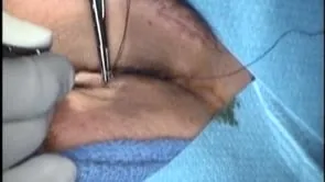
This is Richard Allen at oculosurg.com. This video demonstrates harvesting of a sural nerve graft. The sural nerve provides sensation to the lower lateral leg, lateral heel, ankle, and dorsal foot. The patient will undergo corneal neurotization. The sural nerve is located posterior to the lateral malleolus. The inferior edge of the malleolus has been marked. We will make a straight incision extending from the level of the superior edge of the lateral malleolus superiorly approximately 8 cm. The incision is made midway between the posterior edge of the malleolus and the Achille’s tendon. This incision is often extended in a curvilinear fashion posterior and inferior to the malleolus. A 15 blade is used to make an incision through the skin to the underlying subcutaneous fat. Blunt dissection can then be performed with Metzenbaum scissors. In this area, you will encounter the sural nerve and the small saphenous vein. A superficial vein is encountered initially that can be mistaken for the small saphenous vein. The small saphenous vein runs parallel and in close proximity to the sural nerve. In this case, the small saphenous vein will be posterior to the nerve, but it may be anterior to the nerve. Metzenbaum scissors are used to bluntly dissect in a parallel direction to the path of the sural nerve. The sural nerve and the small saphenous vein are then identified running in close proximity and parallel to each other. The Metzenbaum scissors are then used to sharply and bluntly dissect between the small saphenous vein and the sural nerve. Care is taken not to transect either structure. In this case, a 6 cm length of nerve is needed as the patient will undergo ipsilateral neurotization from the infraorbital nerve. The two structures are dissected from the surrounding tissue to demonstrate the anatomy of the area. Posterior to the small saphenous vein is the Achille’s tendon. The nerve is then transected at its proximal end with the Metzenbaum scissors. The nerve is then transected at its distal end. The distal end is marked for orientation. Measuring the graft shows that it is approximately 6 cm. Inspecting the harvest site shows the small saphenous vein and Achille’s tendon. The incision can then be closed with deep interrupted 4-0 Vicryl or Monocryl sutures and interrupted 5-0 Prolene sutures.