Removal of Medial Orbital Wall and Orbital Floor Implant
Transcript
It is very rare that you can save an infected implant. In general, infected foreign bodies need to be removed. In this video, the patient has a history of multiple recurrent infections of an implant that covers the orbital floor and medial wall. Although responsive to antibiotics, the infection recurs after discontinuation. I rarely will place another implant at the time of implant removal. I might place autologous tissue if I really need support. I believe that the area is contaminated, and that the risk is significant for infection of the new foreign body. I will let the patient heal, and I have been amazed at how many times an additional implant is not needed, as fibrosis/scar tissue adds enough support to prevent any motility deficit or enophthalmos.
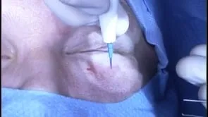
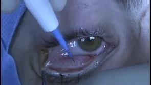
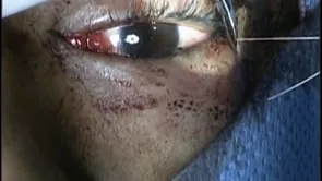
This is Richard Allen at oculosurg.com. This video demonstrates removal of an implant with a history of recurrent infections. The implant covers the medial orbital wall and orbital floor. On imaging, there is no evidence that a fixation screw was used. A 4-0 silk suture is placed through the lower eyelid at the level of the tarsus to provide traction during the case. The needle tip cautery is used to make an incision inferior to the inferior boarder of the tarsus extending from the level of the punctum medially to the lateral canthus laterally. Dissection is the carried out inferiorly between the orbital septum and the orbicularis muscle to the inferior orbital rim. 4-0 silk suture is placed through the cut edge of the conjunctiva/lower lid retractors/orbital septum to provide traction. The inferior orbital rim is exposed. The needle tip cautery is used to make an incision through the periosteum of the inferior orbital rim. The freer periosteal elevator is used to elevate the periosteum from the inferior orbital rim and orbital floor. The implant is identified on the inferior orbital floor and the freer periosteal elevator is used to dissect on top of the implant. This is performed to identify the lateral edge of the implant. Dissection is then directed beneath the implant. Mobilization of the implant is then performed on top and beneath the implant with the freer and the suction. This is a Medpor implant and there is some tissue adherence to the implant that needs to be released. Once the implant appears to be mobilized, a hemostat is used to fixate the implant and slowly remove it from the orbit with a gentle rocking motion. The implant is inspected and will be sent for cultures and sensitivities. The area is inspected for any bleeding and residual foreign body. The conjunctiva is the closed with 7-0 Vicryl suture by recessing the conjunctiva 1-2 mm inferior to the inferior boarder of the tarsus. Antibiotic ointment is placed into the eye, and the patient will follow up in approximately one week.