Radiographic and Clinical Correlation of Microphthalmos with Cyst
Transcript
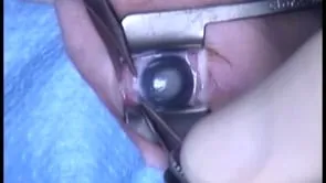
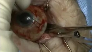
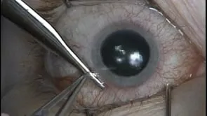
This is Richard Allen at oculosurg.com. This video demonstrates radiographic and clinical correlation of microphthalmos with cyst. In the axial T2 weighted MRI, the microphthalmic globe is noted with a cystic lesion posterior to the globe. The cyst appears to be connected to the globe and optic nerve. On the coronal cut, the cyst is noted to be inferior medial to the optic nerve. The patient underwent enucleation due to inability to wear a scleral shell over the microphthalmic globe. The rectus muscles have been disinserted and the optic nerve will be transected from a lateral approach. In palpating the optic nerve with the scissors, the cyst can be detected and the scissors are directed posterior to the cyst. The optic nerve is transected and the cyst is noted. Examination of the cyst shows its close proximity to the optic nerve and posterior surface of the globe. These cysts are secondary to a defect in closure of the optic fissure which results in extension of a neuroectoderm-lined mass through a coloboma. The cyst often communicates with the subarachnoid space.