Exposure of the Infraorbital Nerve
Transcript
Exposure of the infraorbital nerve can be performed through a transcutaneous or transconjunctival incision. I prefer a transconjunctival incision. This procedure is useful when performing corneal neurotization, or when a biopsy of the infraorbital nerve is needed as in cases of suspected perineural invasion of cutaneous squamous cell carcinoma, or in cases of enlargement of the infraorbital nerve as is seen in IgG4-related disease. It is always important to warn the patient that they will have loss of sensation in the area of the innervation of the infraorbital nerve after then procedure, and that this can be permanent.
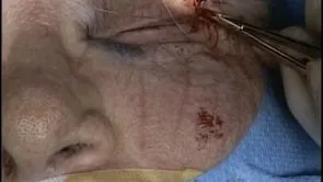
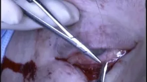
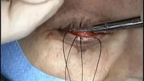
This is Richard Allen at oculosurg.com. This video demonstrates exposure of the infraorbital nerve. This is useful in performing biopsy of the infraorbital nerve and in performing corneal neurotization. A lateral canthotomy and cantholysis has been performed in this case. A transconjunctival incision is made inferior to the inferior border of the tarsus extending from the level of the punctum medially to the lateral canthotomy incision laterally. Dissection is then carried out between the orbital septum and orbicularis muscle to the inferior orbital rim. The inferior orbital rim is exposed and the periosteum of the inferior orbital rim is incised with the needle tip cautery. The periosteum is then elevated from the orbital floor with the Freer periosteal elevator. Dissection does not need to be performed too far posteriorly. The orbital floor is inspected and sometimes the infraorbital nerve can be visualized. If not, the bone of the orbital floor can be palpated at the area of the zygomaticomaxillary suture, where the infraorbital groove should be located. Anteriorly, the infraorbital groove turns into the infraorbital canal. The bone over the infraorbital nerve should be thin and can be fractured easily. This is performed with the suction tip. A Kerrison rongeur can then be used to remove the thin bone superior to the infraorbital nerve. Associated with the infraorbital nerve are the infraorbital vein and artery. Care is taken to just remove bone, without engaging any of the soft tissue with the rongeur. A Freer periosteal elevator can then be used to mobilize the infraorbital nerve from the infraorbital groove and canal.