Lateral Orbitotomy for Cavernous Malformation
Transcript
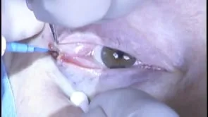
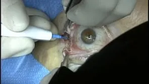
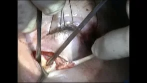
This is Richard Allen in Houston, TX. This video demonstrates a lateral orbitotomy for a cavernous malformation, as we see in the cuts. This mass is lateral to the optic nerve. We could do either an eyelid crease incision or a lateral canthotomy incision. We are doing a lateral canthotomy incision with a lower cantholysis and a superior cantholysis. We will then dissect down to the lateral orbital rim after placing 4-0 traction sutures. The needle tip cautery is used to incise the periosteum of the lateral orbital rim. A freer periosteal elevator is used to elevate the periosteum from the lateral orbital wall. This is usually an avascular plan, but you may run into the zygomaticofacial or zygomaticotemporal neurovascular bundle. The suction can also be used to elevate the periosteum. Malleable retractor is used to protect the globe and the orbit. After adequate elevation has been performed, the periosteum is incised superior the lateral rectus muscle. As we saw in the scan, the mass was relatively superior. Malleable retractors are used to bluntly dissect through the orbital fat. I think this is the most difficult part of the orbitotomy is trying to find the mass. This is in real time, and we are just bluntly dissecting through the orbital fat until we feel or expose the tumor. There it is, there is that cavernous malformation that just popped into view. We will place malleable retractors on either side and then use Takahashi forceps to gently pull on the mass to see if it is mobile. This one is going to prolapse forward so I feel comfortable in pulling on it. We place a 4 X 4 to help with hemostasis. I do not feel we to use cautery. Measuring the mass shows that it is approximately 1.5 cm in diameter. We remove the 4-0 silk sutures. The lateral cantholysis will then be repaired with a 4-0 Vicryl suture which engages the periosteum followed by the periorbita followed by the lateral upper and lateral lower eyelid at the level of the tarsus. This suture is then tied and the level of the lateral canthus is examined to make sure we are in appropriate position. The lateral canthotomy can then be simply closed with 5-0 fast absorbing sutures.