Corneal Neurotization
Transcript
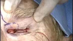
Surgery begins with a subrow incision. Next to section through the orbicularis is performed with Stevens scissors. This allows access to the orbital rim. The arcus marginality is then revealed and the supraorbital notch is palpated. Next, a section through septum is performed superior medially in the area of the supraorbital notch to reveal the supraorbital nerve. Here you can See the lateral aspect of the supraorbital nerve. It's important to avoid the supraorbital artery. Next dissection of the perineurium is performed. This Allows for a bear area for the end to side anastomosis. Next, a S fornix tunnel is made to the orbital dissection and a silk guide suture is placed. This allows for an a traumatic passage of the donor nerve graft through the tunnel. The silk suture is lassoed around the donor nerve graft and the graft advanced through the tunnel. Here we're using a donor cadaver nerve graft. This one is a 50 by three millimeter graft, and you can use other nerve graft such as a sal nerve graft from the patient. However, using a donor nerve graft decreases donor site morbidity. You can see the nerve graft being measured and drawn through, and then end to side anastomosis performed of the donor nerve graft to the supraorbital nerve. Approximately three to four 10 oh nylon sutures are placed to achieve and to side anastomosis. Amniotic membrane graft is then placed over this anastomosis. Next Mies are performed. This is achieved here with a permia at four o'clock as well as eight o'clock. Now some surgeons use large peries, almost 180, but we are choosing to use smaller perms to try to achieve improved healing. You can see here wescott scissors are being used to create a sub tenons tunnel to the area where the donor nerve graft has been drawn through the superior Forex. This is performed at both the four o'clock and the eight o'clock positions to create tunnels where the two main sles will be drawn through in order to provide innervation to the cornea. Here, the sub tenons pocket is being completed from the eight o'clock position so that the sles can be drawn through next. The donor perineurium is dissected. The whole sheath needs to be dissected until the end of the fales and then divided into two main fales. Each of these main fas are then divided into four to eight sles. When we do this division, it's important not to damage the nerve fibers. Then the sles are drawn through the sub tenons tunnels. This is the one for the eight o'clock position, and you can see all the different divided sles when it's drawn through and through four o'clock. The fascicle are then trimmed and drawn to six o'clock and fibrin glue closure performed. Amniotic membrane graft is then placed over the cornea. The subrow incision is then closed and a torso, feet placed, and the patient is patched for one week.