Repair of Full-thickness Upper Eyelid Defect
Transcript
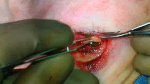
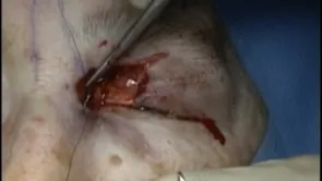
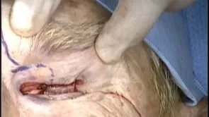
This is Richard Allen at the University of Iowa. This video demonstrates repair of an upper eyelid full thickness defect with a combination of direct closure and a myocutaneous flap. Inspection of the area shows the nature of the defect. Transposition of the eyelid into position shows a bit of redundancy of the anterior lamella compared to the posterior lamella. Therefore, an incision will be made along the eyelid crease to the lateral aspect of the defect. Dissection will then be carried out between the orbicularis muscle and the orbital septum superiorly. This will result in undermining and mobilization of the flap. This will ultimately allow redistribution of the anterior lamella superiorly along the eyelid crease.
This is performed along the full extent of the defect in between the orbicularis muscle and the orbital septum. A small pedicle of tissue is excised to smooth out the line of the flap. Attention is then directed to the eyelid margin which can be repaired primarily. Placement of the anterior lamella superiorly shows that it can be redistributed well along the repair. The eyelid margin will be closed by placing five sero vicral sutures through the anterior surface of the Tarsus. Two sutures will be placed and tied. The lid margin will then be repaired with seven dash zero vicral suture, which will be placed at the level of the meibomian gland orifices. This is a vertical mattress suture which will evert the eyelid margin to prevent postoperative notch. An additional vertical mattress suture will then be placed at the level of the lash follicles and the anterior lamella is then redistributed along the eyelid crease. This will be closed with the same seven dashs vicral suture a success. Zero monofilament suture could be used if preferred. At the conclusion of the case, the defect is repaired without creating an eyelid malposition antibiotic ointment is placed over the area three times a day and the patient will return in one week for a reevaluation and suture removal.