Repair of Lower Lid/Cheek Defect with a Full-thickness Skin Graft
Transcript
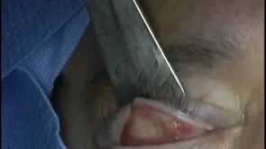
This is Richard Allen at the University of lowa. This video demonstrates placement of a skin graft to a relatively large lower eyelid slash cheek defect. The patient also has involvement of the lateral lower eyelid. The skin in this patient is relatively tight due to actinic damage.
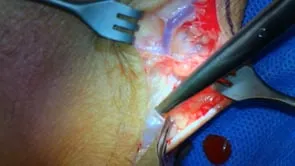
Therefore, a regional flap would be difficult. A perio strip will be raised from the lateral orbital rim to cover the posterior LA mela of the lower lid defect. This is in size with the 15 blade followed by elevation with a freer perio elevator. Inspection of the defect shows that it should be able
to be covered with the periosteal flop. The anterior lamella is excised from the most lateral portion of the Tarsus Residual Tarsus is removed. The periosteal strip and the lateral tarsus are then opposed. A five dash zero vicral suture is then placed in a mattress fashion.
To overlap the periosteal strip onto the lateral tarsus. This completes the repair of the lower lid posterior lamella. A small flap will be raised laterally in order to make the anterior lamellar defect. Smaller, wide undermining is performed with the needle tip cautery around the defect.
Again, the skin is relatively tight and the complete musta flap will not be performed. The posterior surface of the flap is engaged with four deaths, biral suture, which then engages the periosteum of the lateral orbital rim. The suture is then tied which places the flap into position.
The small superior medial portion of the flap is suture to cover the periosteal strip. The closure of the flap is then performed with deep interrupted four deser vicral sutures. Superficial. Five desal proline sutures are then placed on the skin. A template of the defect is made in this case. A supraclavicular graft was harvested. This is sutured into position with five deaths, fast absorbing sutures. These are placed in a running and interrupted fashion. Six. Death sero silk sutures will then be placed around the graft so that the bolster can be fixated. Erything Synap found ointment is placed over the graft. A bolster of telfa with a surgical sponge is then placed into position. The patient will be patched. The patient will follow up in approximately one week for patch removal and reevaluation.